Medical Imaging and Signal Processing Teaching Module
Labs created for Applied Machine Learning EEC174AY in Medical Imaging and Signal Processing
This page highlights two mini-projects personally created and developed for college-level machine learning students. These projects focus on real-world applications of deep learning techniques in biomedical signal processing and medical imaging. All solutions and outputs shown here are generated by me as the class is actively using this lab.
Overview of Mini-Projects
These projects demonstrate the following skills:
- Biomedical Signal Processing: Preprocessing and analyzing ECG signals.
- Custom Neural Network Design: Designing a 1D CNN for signal classification.
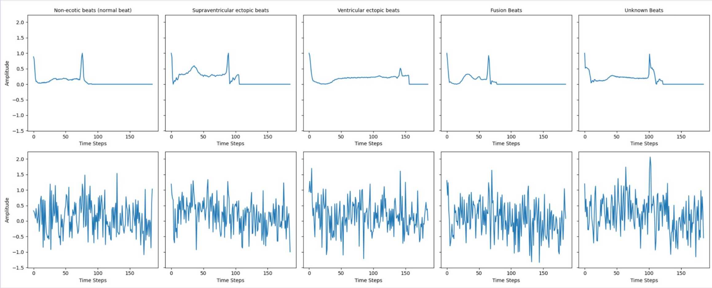
- Data Augmentation and Visualization: Enhancing datasets and interpreting waveforms.
- Deep Learning for Medical Imaging: Semantic segmentation on lung CT scans using pre-trained models.
- Training and Evaluation: Generating learning curves, confusion matrices, and medical statistics.
Explore the assignment notebooks here:
Phase 1: Arrhythmia Classification Using 1D CNN
Phase 2: Lung CT Scan Segmentation
Mini-Project 1: Arrhythmia Classification Using 1D CNN
Objective
To classify different types of arrhythmias from ECG signals using a custom-designed 1D CNN, named ECGNet, on the MIT-BIH dataset.
Dataset Information
- Number of Samples: 109,446
- Categories: 5
- Non-ectopic beats (normal)
- Supraventricular ectopic beats
- Ventricular ectopic beats
- Fusion beats
- Unknown beats
- Resampling: The dataset was resampled to balance class distribution.
Skills and Learning Outcomes
- Designing and training a custom 1D CNN for biomedical signal classification.
- Visualizing data augmentation and performance metrics.
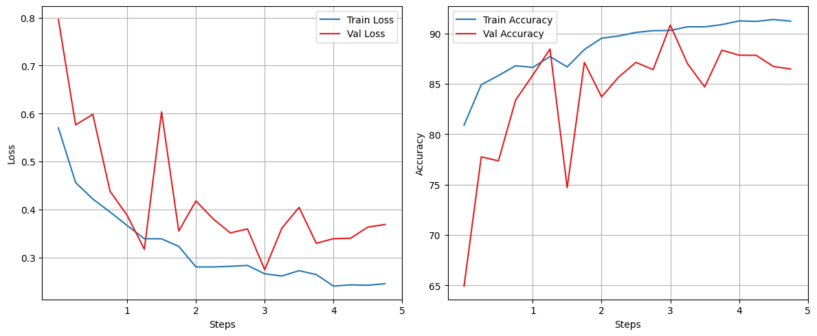
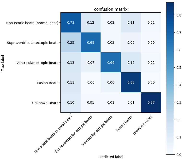
- Evaluating models using learning curves and confusion matrices.
Sample Outputs
Note: The following outputs are examples generated by me while creating the lab and solutions. Solutions and full implementations are withheld to maintain academic integrity.
- Waveform Visualization Before and After Augmentation

- Model Evaluation Metrics


Mini-Project 2: Lung CT Scan Segmentation
Objective
To segment lung regions from CT scans using a pre-trained ResNet34-based U-Net architecture.
Dataset Information
- Number of Scans:
- 266 2D scans with masks
- 4 3D scans with masks
- Supplementary Data: Includes
lung_stats.csvfor statistical analysis.
Skills and Learning Outcomes
- Applying semantic segmentation techniques using pre-trained models.
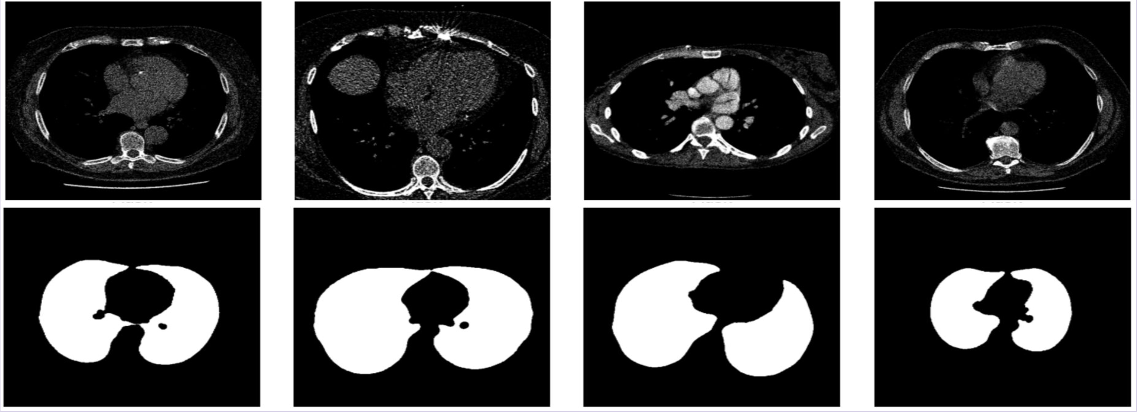
- Visualizing medical imaging data and segmentation results.
- Extracting and analyzing medical statistics from segmentation predictions.
Sample Outputs
Note: The outputs below are sample results generated by me during the lab creation process. Solutions and full implementations are withheld to maintain academic integrity.
- Ground Truth Masks Visualization

- Segmentation Results

- Statistical Analysis Results

Key Takeaways
These projects illustrate:
- Hands-on experience in designing and evaluating deep learning models for healthcare applications.
- Application of 1D CNNs and semantic segmentation models for biomedical data.
- Preparing students for challenges in Health AI and medical image analysis, equipping them with research and industry-relevant skills.
Acknowledgment
The GIF used as the cover for the lung segmentation project is sourced from Imbio’s Lung Density Analysis. All rights belong to Imbio.